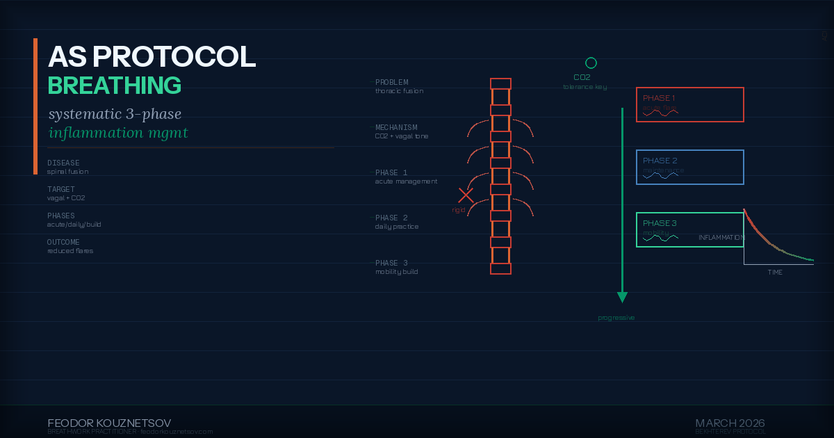
Ankylosing Spondylitis Breathing Protocol — Systematic Approach to Managing Inflammation, Mobility, and Pain Through Respiratory Function
Why Ankylosing Spondylitis Requires a Specific Breathing Protocol
Ankylosing spondylitis (AS) is a chronic inflammatory disease that primarily affects the spine and sacroiliac joints. Over time, inflammation can cause vertebrae to fuse, reducing spinal mobility and creating progressive rigidity. But the respiratory implications are often overlooked: AS affects the thoracic spine and rib cage, which directly impairs breathing mechanics.
As the disease progresses, the rib cage becomes less mobile. Costovertebral joints (where ribs attach to vertebrae) calcify and fuse. This reduces thoracic expansion during inhalation, forcing the diaphragm to compensate. Many people with AS develop shallow, rapid breathing patterns as the chest wall becomes increasingly rigid. This creates a feedback loop: restricted breathing → increased sympathetic activation → more inflammation → more pain → more breathing restriction.
I have lived with AS since diagnosis in 2009 and have managed the condition without constant medication since 2015 using breathing protocols as the primary intervention. This is covered in detail in the AS management page and personal story post. This post provides the systematic protocol — the specific techniques, phases, and progressions that address AS symptoms through respiratory function.
Understanding AS Respiratory Dysfunction
Thoracic Restriction and Breathing Mechanics
Normal breathing involves coordinated movement of the diaphragm, intercostal muscles, and chest wall. The diaphragm contracts and descends during inhalation, creating negative pressure that draws air into the lungs. The ribs expand laterally and the sternum lifts anteriorly, increasing thoracic volume. This is three-dimensional expansion: vertical (diaphragm), lateral (ribs), and anterior (sternum).
AS impairs all three dimensions. Vertebral fusion reduces spinal extension and lateral flexion. Rib cage calcification limits lateral expansion. Anterior chest wall rigidity restricts sternal lift. The result: breathing becomes predominantly diaphragmatic because thoracic expansion is mechanically blocked.
This is not inherently problematic — diaphragmatic breathing is more efficient than chest breathing. But when the thoracic component is lost entirely, several issues develop:
- Reduced lung capacity: Maximum inhalation volume decreases because thoracic expansion is restricted
- Increased breathing rate: To maintain adequate ventilation with reduced tidal volume, breathing frequency increases
- Hyperventilation: Faster breathing reduces CO2, which increases inflammation and pain sensitivity (covered in the hyperventilation post)
- Postural compensation: Forward head posture and rounded shoulders develop to maximize remaining lung capacity, which worsens spinal alignment
Inflammation and CO2
Chronic hyperventilation reduces blood CO2 levels. Low CO2 shifts the body toward a more alkaline pH (respiratory alkalosis), which has been shown to increase inflammatory cytokine production and reduce tissue oxygenation via the Bohr Effect (covered in the Bohr Effect post).
In AS, this creates a direct feedback loop: inflammation → pain → shallow rapid breathing → low CO2 → more inflammation → more pain. Breaking this loop requires deliberately increasing CO2 tolerance through breathing retraining. This is the foundation of the protocol.
The Protocol: Three-Phase Approach
The AS breathing protocol is organized into three phases based on disease activity and symptom severity. Each phase has specific goals and techniques. Progression between phases is not linear — you move between phases based on current inflammation levels, not calendar time.
Phase 1: Acute Flare Management (High Inflammation, High Pain)
Goal: Reduce sympathetic activation and pain sensitivity without increasing mechanical stress on inflamed joints
Duration: Used during active flares until pain and stiffness decrease to manageable levels (typically 3-10 days)
Techniques:
1. Extended Exhalation (4:8 Ratio)
- Inhale through nose for 4 seconds (minimal chest expansion, primarily diaphragmatic)
- Exhale through nose for 8 seconds (slow, controlled, no force)
- Repeat for 5-10 minutes, 2-3 times daily
- Practice lying supine or semi-reclined to minimize postural stress
Mechanism: Extended exhalation activates vagal efferents, shifting autonomic state toward parasympathetic dominance. This reduces inflammatory cytokine production and lowers pain sensitivity without requiring joint movement.
2. Coherent Breathing (5-6 Breaths Per Minute)
- Inhale through nose for 5 seconds
- Exhale through nose for 5 seconds
- Maintain smooth, even rhythm with no breath holds
- Practice for 10-20 minutes, 2 times daily
Mechanism: Breathing at resonance frequency (5-6 breaths per minute) maximizes heart rate variability (HRV) and strengthens vagal tone. Covered in detail in the coherent breathing post. This is the most effective technique for reducing baseline inflammation during flares.
3. Humming (Vagal Activation + Nitric Oxide)
- Inhale through nose for 4 seconds
- Hum on exhalation for 6-8 seconds (comfortable pitch, moderate volume)
- Repeat for 5 minutes, 1-2 times daily
Mechanism: Humming stimulates mechanoreceptors in the nasal passages and larynx, activating vagal afferents. It also increases nitric oxide (NO) production in the paranasal sinuses. NO has anti-inflammatory effects and improves blood flow, which supports tissue healing during flares.
Phase 1 Protocol Summary: During acute flares, practice extended exhalation upon waking (5-10 min), coherent breathing midday (10-20 min), and humming before sleep (5 min). Total daily practice: 20-35 minutes. No mobility work, no breath holds, no forceful breathing — only gentle regulation to reduce inflammation and pain.
Phase 2: Baseline Maintenance (Low-Moderate Inflammation, Manageable Pain)
Goal: Maintain CO2 tolerance, preserve remaining thoracic mobility, prevent progression of breathing pattern dysfunction
Duration: Used when not in active flare — this is the default protocol for daily practice
Techniques:
1. Buteyko Breath Holds (CO2 Tolerance Training)
- Sit comfortably, breathe normally through nose for 1 minute
- After a normal exhalation, pinch nose and hold breath
- Hold until moderate air hunger (not distress — stop before gasping)
- Release and resume nasal breathing (slowly, not gasping)
- Wait 1 minute, repeat for 4-5 rounds
- Practice once daily, preferably morning
Mechanism: Breath holding increases CO2 levels, which recalibrates chemoreceptors to tolerate higher CO2. This reduces baseline breathing rate and prevents hyperventilation. Critical for breaking the inflammation-hyperventilation loop. Covered in the Buteyko mechanism post.
2. Thoracic Mobility Breathing (Rib Cage Expansion)
- Stand or sit upright, hands on lower ribs (lateral position)
- Inhale slowly through nose, focus on expanding ribs laterally (pushing hands outward)
- Exhale slowly, allow ribs to return to neutral (no force)
- Repeat for 10 breaths, 2 times daily
Mechanism: Deliberate lateral rib expansion maintains remaining mobility in costovertebral joints. This is not stretching — it is active movement within current range of motion to prevent further restriction. Even minimal improvement in thoracic expansion increases lung capacity and reduces compensatory breathing patterns.
3. Nasal Breathing During All Activity
- Maintain nasal breathing during walking, daily tasks, and light exercise
- If air hunger forces mouth breathing, slow down until nasal breathing is sustainable
- Use mouth taping during sleep (if nasal breathing is functional at rest)
Mechanism: Nasal breathing maintains CO2 levels during activity, prevents exercise-induced hyperventilation, and delivers continuous nitric oxide. This is covered in the exercise breathing post. For AS, this prevents flare triggers caused by activity-induced hyperventilation.
Phase 2 Protocol Summary: Practice Buteyko breath holds in morning (5 min), thoracic mobility breathing midday and evening (2 min each), coherent breathing before sleep (10 min). Maintain nasal breathing during all waking activity. Total daily practice: 20 minutes formal + continuous nasal breathing.
Phase 3: Mobility and Capacity Building (Minimal Inflammation, Stable Pain)
Goal: Maximize remaining respiratory capacity, build exercise tolerance, prevent deconditioning
Duration: Used when baseline inflammation is well-controlled and pain is minimal — typically after months of consistent Phase 2 practice
Techniques:
1. Breath Hold Walking (Progressive CO2 Challenge)
- Walk at comfortable pace, breathing normally through nose
- After normal exhalation, hold breath and continue walking
- Hold until strong air hunger, then resume nasal breathing
- Walk normally for 1-2 minutes, repeat
- Practice 3-5 rounds during daily walk
Mechanism: Combining breath holds with movement creates a stronger CO2 stimulus than breath holds at rest. This builds deeper CO2 tolerance and trains the body to maintain nasal breathing under metabolic stress. Essential for preventing hyperventilation during physical activity.
2. Thoracic Extension Breathing
- Stand with hands clasped behind lower back
- Gently lift chest and extend thoracic spine (within comfortable range)
- Inhale deeply through nose, focusing on anterior chest expansion
- Hold for 2-3 seconds at full inhalation
- Exhale slowly and return to neutral
- Repeat for 5-8 breaths, once daily
Mechanism: Thoracic extension combined with deep inhalation mobilizes the anterior chest wall and strengthens remaining intercostal function. This counteracts the forward flexion tendency in AS and maintains maximum available lung capacity.
3. Exercise Integration (Nasal Breathing Under Load)
- Walking, swimming, cycling, or other low-impact aerobic activity
- Maintain nasal breathing throughout entire session
- If intensity forces mouth breathing, reduce intensity until nasal breathing is sustainable
- Gradually increase duration and intensity while maintaining nasal breathing
- Practice 3-5 times per week, 20-40 minutes per session
Mechanism: Regular aerobic exercise with nasal breathing maintains cardiovascular fitness, prevents deconditioning, and builds CO2 tolerance under functional load. Swimming is particularly beneficial for AS because horizontal position reduces spinal compression and water provides gentle resistance that supports thoracic mobility.
Phase 3 Protocol Summary: Morning breath hold walking (10 min), thoracic extension breathing midday (3 min), nasal breathing exercise 3-5x weekly (20-40 min), coherent breathing before sleep (10 min). Total daily practice: 25 minutes formal + 60-120 minutes weekly exercise.
Measurable Outcomes and Progression
Track these markers to assess protocol effectiveness and guide phase progression:
- Control Pause (CP): Buteyko measurement of CO2 tolerance. Exhale normally, hold breath, time until first urge to breathe. Target: 30+ seconds indicates good CO2 tolerance. Track weekly.
- Resting breathing rate: Count breaths per minute at rest. Target: 10-12 breaths/min. Higher rates indicate hyperventilation. Track daily for first month, then weekly.
- Chest expansion: Measure chest circumference at full inhalation and full exhalation. Difference indicates thoracic mobility. Target: maintain or increase range. Track monthly.
- Pain levels: Subjective 0-10 scale. Track daily. Look for reduction in baseline pain and faster recovery from flares.
- Morning stiffness duration: Minutes from waking until full mobility restored. Track daily. Target: reduction over months.
- CRP/ESR (if monitored): Inflammatory markers from blood tests. Track per medical schedule. Look for reduction correlating with breathing practice.
Progression between phases is based on symptoms, not time. Return to Phase 1 during flares regardless of how long you have been practicing Phase 2 or 3. The protocol is flexible and responsive to disease activity.
Integration with Medical Treatment
This breathing protocol is not a replacement for medical treatment. AS requires medical monitoring and, for many people, pharmaceutical intervention. The protocol is complementary:
- NSAIDs: Breathing practice may reduce need for constant NSAID use by lowering baseline inflammation, but this should be discussed with your rheumatologist
- Biologics: If prescribed, continue as directed. Breathing practice supports but does not replace biologic therapy
- Physical therapy: Breathing mobility work complements PT exercises for spinal mobility and posture
- Regular monitoring: Continue regular rheumatology appointments and inflammatory marker testing
For my personal management, I transitioned off NSAIDs in 2015 after two years of consistent breathing practice combined with anti-inflammatory nutrition and stress management. This was done gradually under medical supervision with regular CRP monitoring. Not everyone will have this outcome, and it should never be attempted without medical guidance.
Common Challenges and Troubleshooting
Challenge: Air Hunger During Breath Holds
Solution: Start with shorter holds (10-15 seconds) and progress gradually. Air hunger is the point of the practice — it signals rising CO2, which is what you are training tolerance to. Discomfort is expected. Panic or distress is not. If breath holds trigger anxiety, practice extended exhalation instead until CO2 tolerance improves.
Challenge: Thoracic Expansion Causes Pain
Solution: Reduce range of motion. The goal is movement within pain-free range, not maximal stretch. Even 1-2mm of rib expansion is beneficial. If all thoracic movement is painful, return to Phase 1 techniques (no mobility work) until inflammation reduces.
Challenge: Cannot Maintain Nasal Breathing During Exercise
Solution: Reduce intensity. If you cannot sustain nasal breathing at a given exercise intensity, your CO2 tolerance is not yet sufficient for that load. Slow down until nasal breathing is comfortable, then gradually increase pace over weeks. This is covered in detail in the exercise breathing post.
Challenge: Flares Occur Despite Consistent Practice
Solution: Flares are part of AS. Breathing practice reduces frequency and severity but does not eliminate them. During flares, shift immediately to Phase 1 protocol. Do not attempt mobility work or breath holds when inflammation is high. The goal is management, not cure.
When Individual Guidance Helps
For people with AS who are new to breathing practice or who have significant thoracic restriction, individual work helps establish the protocol correctly and troubleshoot specific challenges. The assessment identifies baseline respiratory function, CO2 tolerance, and thoracic mobility. And in individual sessions, I guide technique progressions tailored to your specific AS presentation and current disease activity.
This protocol is based on my 11 years of managing AS without constant medication. It works. But it requires consistency, patience, and proper progression. The breathing pattern dysfunction that develops with AS does not reverse in weeks — it shifts gradually over months with daily practice. This is not quick relief. It is systematic retraining that addresses the underlying respiratory and autonomic dysfunction maintaining the inflammatory cycle.
Next step
Want to work through your situation personally?
Disclaimer: personal experience & self-regulation practices. Not medical advice.